Preparing an AMBER topology file for a protein plus ligand system: Difference between revisions
import>Em427 No edit summary |
import>Em427 No edit summary |
||
| Line 94: | Line 94: | ||
WILL FINISH TOMORROW |
WILL FINISH TOMORROW |
||
do not forget about Ca->Mg and CD1->CD for isoleucine :) |
|||
Revision as of 18:12, 12 May 2008
When you want to use AMBER, either on its own to run MD, or interfaced with any of the group software (GMIN, OPTIM or PATHSAMPLE), you need to have two files at your disposal. These are the topology file (which describes the atom types, connectivity of atoms, force field parameters for angles, etc.) and the coordinate file (which shockingly defines the coordinates of the atoms in your system).
For big systems, generating these files can take a bit of time, and the difficulty increases when you are trying to simulate something for which parameters do not exist in the AMBER libraries i.e. non-standard amino acids, or, more usually, a system with a novel ligand (for example a drug molecule). This tutorial will deal with a big system, hopefully covering all bases. You will probably find the process much less arduous when you are dealing with something smaller and I will try to point out where I am deviating from what you would need to do if you only had say a protein composed of standard amino acids.
I will also assume that you have already generated parameters (AMBER prepin and frcmod files) for any novel ligand/molecule that is present in your system. Info on how to do this can be found linked from the AMBER page.
Right! Lets begin:
Step 1: Preparing your pdb file
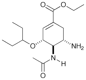
For this tutorial, I will be looking at neuraminidase (NA), an Influenza virus surface protein whose natural substrate is sialic acid. There are a few drugs which target this molecule as competitive inhibitors and one well known example is Tamiflu. Shown on the right is in fact the pro-drug form which you take. In the body, the ester is hydrolysed to a carboxylic acid and this is the form I will be looking at.
The starting point is a crystal structure of Tamiflu (oseltamivir) bound to NA which I obtained from the Protein Data Bank. NA is actually a tetramer so to cut down the simulation time, I will only consider the monomer and therefore I removed all but one copy from the PDB file, just by deleting the entries for the atoms.
Starting point: 2hu4.pdb
Removing Hydrogens from the protein
As AMBER uses an all-atom representation, we need to allow it to add in Hs as it sees fit when you generate the topology file. This is a problem if you already have Hs in the structure so for any part which you are going to load parameters for from the AMBER libraries, you must remove ALL H. This is best done using a script or a series of piped commands. For example, first, you find which types of H you have in the molecule using:
grep H 2hu4.pdb | more
You'll get something like this:
ATOM      2  H1  VAL     1      43.584  25.333  13.539  1.00  0.00
ATOM      3  H2  VAL     1      44.124  26.003  14.929  1.00  0.00
ATOM      4  H3  VAL     1      42.554  25.613  14.779  1.00  0.00
ATOM     12  H   LYS     2      43.614  29.813  13.829  1.00  0.00
ATOM     19  HZ1 LYS     2      48.594  30.403  12.569  1.00  0.00
ATOM     20  HZ2 LYS     2      47.324  29.383  12.669  1.00  0.00
ATOM     21  HZ3 LYS     2      47.194  30.783  11.829  1.00  0.00
ATOM     25  H   LEU     3      41.774  31.293  17.769  1.00  0.00
ATOM     34  H   ALA     4      38.304  32.193  15.399  1.00  0.00
ATOM     40  H   GLY     5      37.974  36.783  16.069  1.00  0.00
ATOM     45  H   ASN     6      35.304  36.683  15.349  1.00  0.00
ATOM     51 HD21 ASN     6      34.094  36.233  14.939  1.00  0.00
ATOM     52 HD22 ASN     6      33.744  35.023  13.739  1.00  0.00
...
The task now is to find a pattern which will let you remove each type of H in turn using grep -v. i.e. I can remove the first three types of H using:
grep -v ' H[1-3]' 2hu4.pdb
NOTE: I've got a space before the H there. This is so that I don't accidentally remove lines with NH1 on for example. A good method is just to do a normal grep for the pattern that you plan on using to check that you won't catch any atoms you don't intend to! Once you've found a patten that covers all types of H in your molecule, you need to string them together and redirect the output to a new pdb file e.g.
grep -v ' H[1-3] ' 2hu4.pdb | grep -v H[Z,D,H,G] | grep -v ' HE' | grep -v ' H ' > 2hu4_noH.pdb
The results is here: 2hu4_noH.pdb
Don't worry about the atom numbering being messed up, we'll deal with that later! Remember also that command presented above fits to 2hu4_noH.pdb file. Other PDB file can contain Hs with different names.
Adding H to the ligand
In my pdb file, there is a residue OTV corresponding to the drug molecule. When I generated parameters in AMBER for that residue, it had a complete set of Hs, and crystal structure from the PDB doesn't have any more. This is a problem! We will need to add them back in using e.g. Molfacture, a tool in VMD. You will need the latest version of VMD to do this so either download and compile it yourself (which is pretty easy - honest!), or use my binary located at
clust:~csw34/bin/vmd
Using Molfacture to do this is explained in a separate tutorial (as it's a bit long winded) in the VMD section. Once you have added all the Hs required and ensured that the charge on the molecule is what you require (-1 in this case due to a COO- group), you need to save the ligand you are editing as a new PDB using File>Save and changing the type to .pdb. The result is linked below:
Ligand plus Hs from VMD: vmdligand.pdb
NOTE: The coordinates have been preserved from the original crystal structure position of the ligand. This will turn out to be very useful later on!
Checking for consistent atom naming
This is a vital step as when you parametrised your ligand originally, each atom was assigned a name from the generalised forcefield. Unless the atoms names in your pdb match with those in the AMBER prepin file, you're not going to be able to make a topology or coordinate file to use with AMBER. Unfortunately, this just requires some manual editing. Here is one suggested method but feel free to change it if you think there is a more logical way forward. You will need a working copy of xleap for this bit. It is part of the AMBER package so if you do NOT have AMBER installed on your workstation, get someone to help you!
Here are the .prepin and .frcmod files for the OTV ligand that I made earlier (This is sounding a bit like Blue Peter!):
- prepin file: OTV.prepin
- frcmod file: OTV.frcmod
The first thing we need to do is create a .pdb from the .prepin file using xleap. Start xleap and type the following:
loadamberprep OTV.prepin
savepdb OTV OTV.pdb
quit
You will now have OTV.pdb in the directory. Looking at is, there is obviously something a bit wrong! Some of the atom names seem to start with numbers! LEaP is a very powerful tool (apparently), but as such - it has so many options and quirks that using it rapidly becomes a bit of a nightmare. In this case, it has messed around with the atom names and so we will need to correct this mistake. Open up OTV.pdb and OTV.prepin in your favourite editor and, atom by atom, ensure that the pdb file has the same atom names as in the prepin. For example, 1HM2 in the pdb file should clearly be HM21 and so on.
WARNING: DO NOT CHANGE THE PREPIN FILE AT ALL! Doing so will cause all sorts of bad things to happen. Only edit the pdb!
The resulting pdb file will look like this: OTV_checked.pdb. When you used Molfacture to add the Hs onto the ligand in the crystal structure, you also assigned them names but unfortunately, they will NOT be the same as those assigned by xleap (i.e. those we just checked from the prepi file) so now we need to compare OTV_checked.pdb with vmdligand.pdb visually. The best option is to open both pdb files in e.g. Pymol and orient them so they look the same, displaying the atom names from the label menu (aligning would be even better but in this case order of atoms in both PDB files is unfortunately different). Now, open vmdligand.pdb and, one atom at a time, check it's name is the same in both structures. If they are not, correct vmdligand.pdb with the right name.
The result is this: vmdligand_correct.pdb
Now, vmdligand_correct.pdb contains not only the correct atom names in the right order but also the correct xyz coordinates from the original crystal structure. We're ready to splice them back into the original pdb!
Creating the input pdb
Open the crystal structure you removed the Hs from earlier ( 2hu4_noH.pdb) and delete all the atoms belonging to the drug molecule. In their place, paste in the atoms directly from vmdligand_correct.pdb! Ensure that you add a TER line between the end of the protein and the start of the drug, and also between the end of the drug and the CA (Calcium) ion. We're now ready to have a go at making the topology file!
At this stage, we're working with: 2hu4_noH_corrected.pdb
Step 2: Making the initial topology file
WILL FINISH TOMORROW
do not forget about Ca->Mg and CD1->CD for isoleucine :)
